Pranav Rane
Deuterium Ion Implantation of Silicon Dioxide
Introduction
Due to the lack of atmosphere on the Moon, the lunar surface is constantly exposed to solar wind irradiation, galactic cosmic radiation, solar energetic particles, and micrometeorite impacts. While these processes are generally considered destructive, some scientists believe that solar wind exposure and micrometeorite impacts significantly contribute to the formation and movement of water on the lunar surface.\(^{1234}\) Water, an essential resource for long-term lunar exploration, has been confirmed to exist on the Moon by orbital spacecraft.\(^5\) However, the formation and precise location of water within lunar material remain not fully understood. It is believed that varying quantities of \(\text{H}_2\), \(\text{H}_{2}\text{O}\), and/or \(\text{OH}\) form within lunar material after exposure to solar wind, which is comprised of high-energy protons, electrons, and trace amounts of heavy ions. In this process, the protons are thought to penetrate and form bonds within the oxygen-rich regolith. Recently, scientists have identified hydrogen-bearing species residing within vesicles in lunar samples\(^6\) (Figure 1). These vesicles have been shown to form approximately 50 nanometers beneath the surface of the lunar regolith. However, the presence of \(\text{H}_{2}\text{O}\) within these vesicles has yet to be confirmed, and the physical processes governing vesicle formation are not entirely understood.
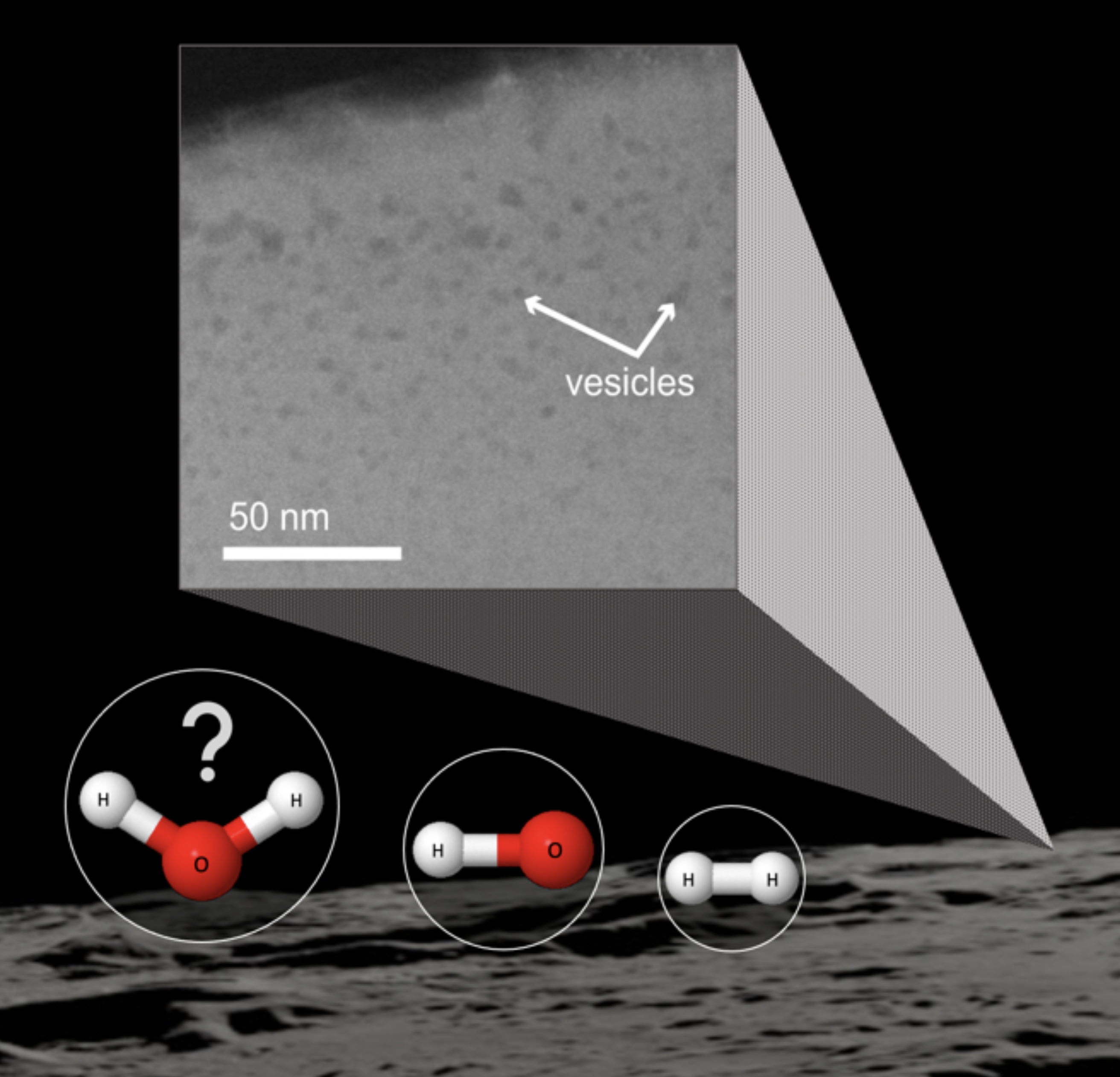
In this study, we perform deuterium ion irradiation on silicon dioxide, a lunar-relevant material, to recreate subsurface vesicles that may contain deuterium-bearing species. We examine the ion-induced damage to these materials to calibrate the deuterium ion source exposure parameters necessary for proper vesicle formation. Our ultimate goal is to use the calibrated deuterium ion source to irradiate Apollo 17 sample 75035.232 and recreate vesicles.
Methods
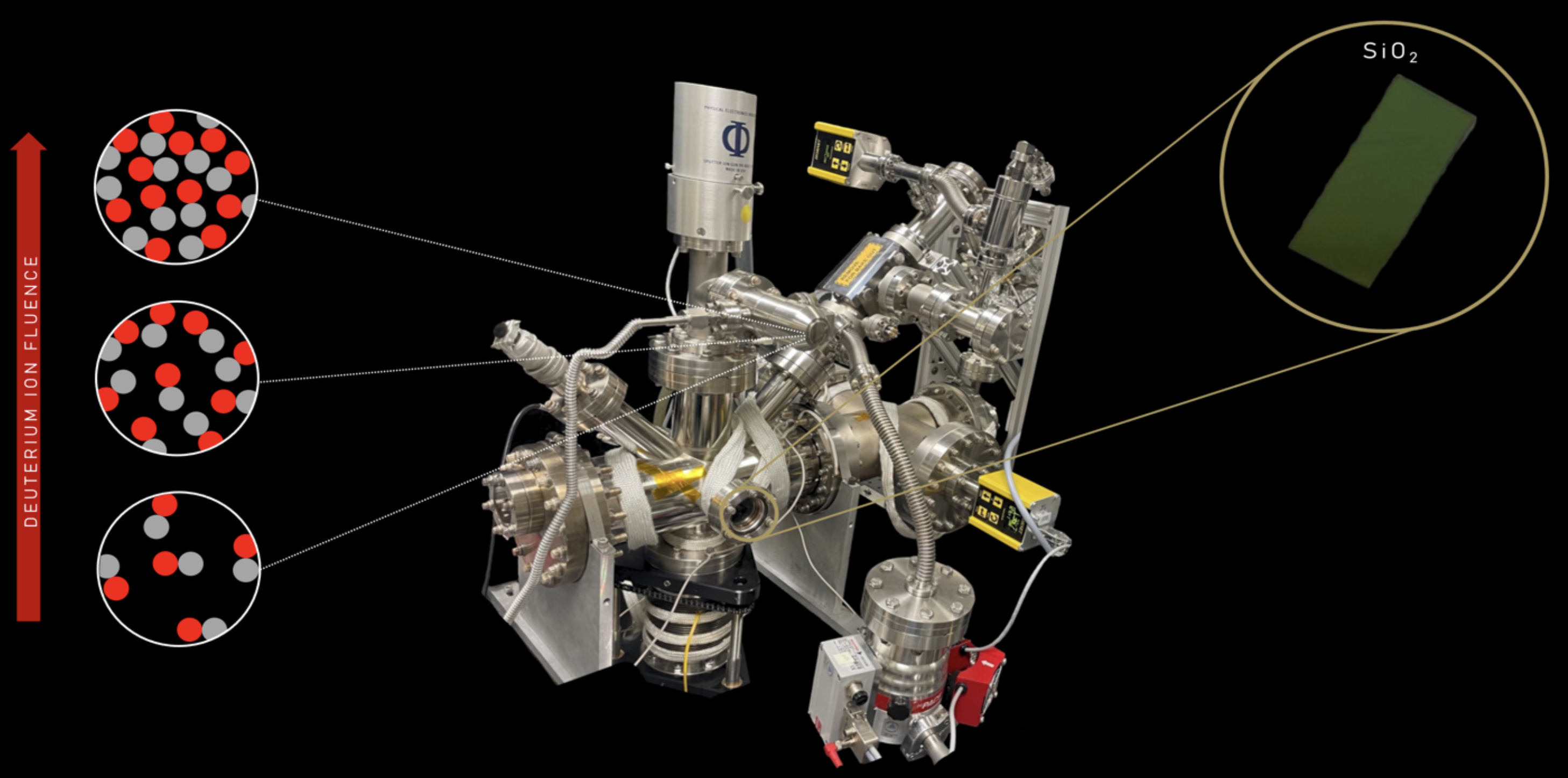
To recreate the conditions of solar wind bombardment, we used a deuterium ion source with variable flux and exposure time (Figure 2). The energy of the deuterium ions is set constant at 2 keV. Additionally, an electron beam was employed to prevent charging during the irradiation of the silicon dioxide samples. The fluence (ions/cm²) of the deuterium source was adjusted by varying the flux and exposure time, facilitating vesicle formation.
During irradiation, we used samples of silicon dioxide with a 300 nanometer oxide layer on a silicon wafer. The samples were exposed to various fluences, and the resulting surface damage was analyzed using atomic force microscopy (AFM) with a Bruker Dimension Icon. Quantitative analysis of blister coverage in silicon dioxide was performed using the StarDist\(^7\) package, which utilizes a neural network pretrained on fluorescent microscopy images of cells. This network effectively computes instance segmentation maps of the deuterium blisters using a star-convex polygon representation.
Results
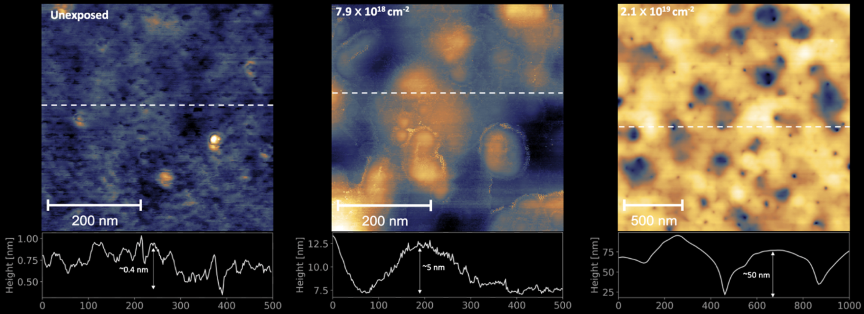
The AFM data of exposed silicon dioxide reveal the creation of blisters on the surface that we believe contain deuterium species. However, at fluences above \(2.1 \times 10^{19}\) \(\text{cm}^{-2}\), the blisters are observed to have burst, resulting in deep cavities on the surface (Figure 3).
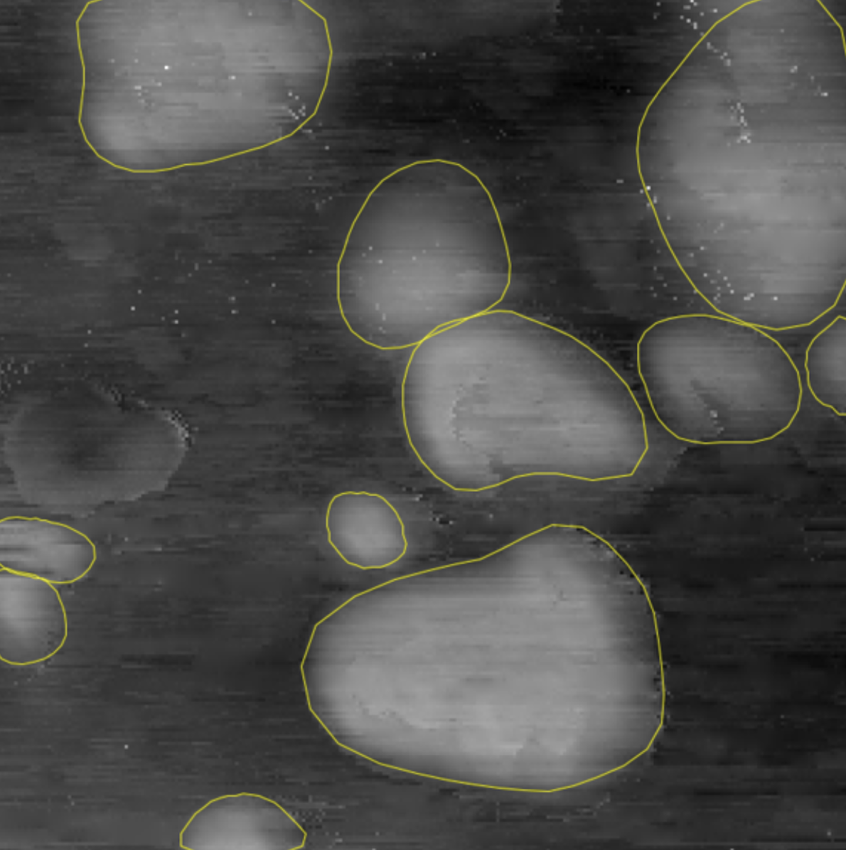
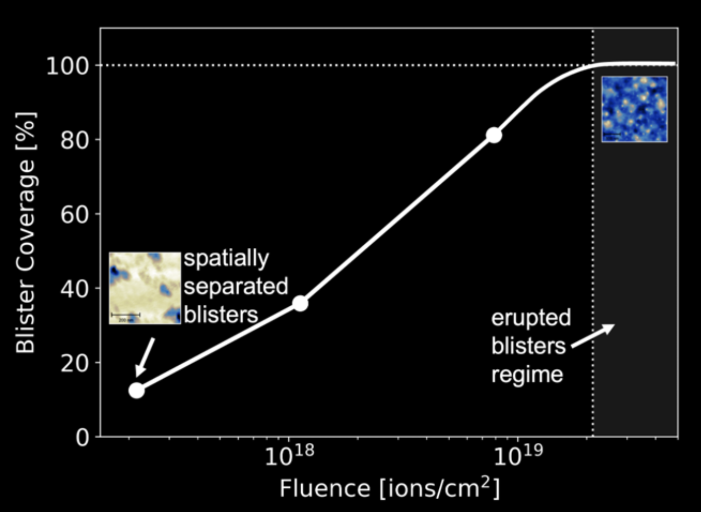
Segmentation results using StarDist on an AFM image of exposed silicon dioxide provide spatial coordinate data for each blister, allowing for the calculation of metrics such as total blister percentage, average blister size, and blister size deviation (Figure 4).
Discussion
In addition to using StarDist, I experimented with another computational approach for segmenting blisters and extracting coordinate data. This method involved a convolutional neural network (CNN) with a modified U-net\(^8\) architecture to perform semantic segmentation. The results produced a binary mask that classified each pixel in the AFM images as either “blister” or “not-blister.” However, we encountered two significant problems with this method.
First, while semantic segmentation effectively classifies pixels into categories, it does not provide a way to extract coordinate data when blisters overlap. Since deuterium blisters begin to overlap at higher fluences (Figure 3), this limitation becomes critical when extracting metrics such as average blister size. Second, the quantity of AFM image data collected was insufficient to train an accurate segmentation algorithm. Although data augmentation can increase dataset sizes, our collected AFM image data was still inadequate. StarDist addresses both issues by using star-convex polygons to represent blisters and leveraging a pretrained neural network. From this, we can accurately extract the average size and density of blisters.
The relationship between ion fluence and blister coverage shown in Figure 5 indicates that a deuterium fluence of less than \(10^{18}\) \(\text{cm}^{-2}\) provides sufficient ion exposure to promote the formation of spatially separated deuterium blisters on the surface without overexposure. We believe these spatially separated deuterium surface blisters indicate the formation of spatially separated subsurface vesicles, as shown in Figure 1.
Our future work includes performing irradiation with the calibrated fluence on anorthite, a lunar-relevant mineral that is more complex than silicon dioxide. We will use focused ion beam (FIB) lift-outs and scanning transmission electron microscopy (STEM) to analyze subsurface vesicle formation in anorthite. Upon confirmation of vesicle formation in anorthite, we will irradiate lunar sample 75035.232 with the same fluence, perform FIB lift-outs and STEM, and analyze any vesicles present. Additionally, we will use nano-FTIR\(^9\) to identify chemical species that may be trapped in vesicles. If \(\text{D}_{2}\text{}\) is found within vesicles in the lunar sample, it would provide concrete evidence that solar wind exposure can facilitate the production of water on the Moon.
References
[1] Jones, B. M., Aleksandrov, A., Hibbitts, K., Dyar, M. D., & Orlando, T. M. (2018). Solar wind‐induced water cycle on the Moon. Geophysical Research Letters, 45(20), 10-959.
[2] Managadze, G. G., Cherepin, V. T., Shkuratov, Y. G., Kolesnik, V. N., & Chumikov, A. E. (2011). Simulating OH/H2O formation by solar wind at the lunar surface. Icarus, 215(1), 449-451.
[3] Bradley, J. P., Ishii, H. A., Gillis-Davis, J. J., Ciston, J., Nielsen, M. H., Bechtel, H. A., & Martin, M. C. (2014). Detection of solar wind-produced water in irradiated rims on silicate minerals. Proceedings of the National Academy of Sciences, 111(5), 1732-1735.
[4] Badyukov, D. D. (2020). Micrometeoroids: the Flux on the Moon and a Source of Volatiles. Solar System Research, 54, 263-274.
[5] Pieters, C. M., Goswami, J. N., Clark, R. N., Annadurai, M., Boardman, J., Buratti, B., ... & Varanasi, P. (2009). Character and spatial distribution of OH/H2O on the surface of the Moon seen by M3 on Chandrayaan- 1. science, 326(5952), 568-572.
[6] Burgess, K. D., Cymes, B. A., & Stroud, R. M. (2023). Hydrogen-bearing vesicles in space weathered lunar calciumphosphates. Communications Earth & Environment, 4(1), 414.
[7] Schmidt, U., Weigert, M., Broaddus, C., & Myers, G. (2018). Cell detection with star-convex polygons. In Medical Image Computing and Computer Assisted Intervention–MICCAI 2018: 21st International Conference, Granada, Spain, September 16-20, 2018, Proceedings, Part II 11 (pp. 265-273). Springer International Publishing.
[8] Ronneberger, O., Fischer, P., & Brox, T. (2015). U-net: Convolutional networks for biomedical image segmentation. In Medical image computing and computer-assisted intervention–MICCAI 2015: 18th international conference, Munich, Germany, October 5-9, 2015, proceedings, part III 18 (pp. 234-241). Springer International Publishing.
[9] Huth, F., Govyadinov, A., Amarie, S., Nuansing, W., Keilmann, F., & Hillenbrand, R. (2012). Nano-FTIR absorption spectroscopy of molecular fingerprints at 20 nm spatial resolution. Nano letters, 12(8), 3973- 3978.